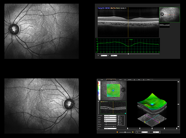
Superb technology leading the way to more definitive diagnosis.
Zeiss OCT Cirrus HD
With world-class ZEISS optics and knowledge from over a decade of experience in OCT, Cirrus captures spectacular
images second to none. Cirrus uses Selective Pixel Profiling & trade; to optimize data at each pixel.
Anterior segment images

Why Should You Care?
-
Spot small areas of pathology. Tightly spaced B-scans ensure that small areas of pathology are imaged. For
reference, a human hair is about 40-120 microns in diameter.
-
Visualize the fovea (center of the macula). Scans that are spaced further apart than in the Cirrus cube may miss
the central fovea.
-
Fuel for analysis. Millions of data points from the cube are fed into the ZEISS proprietary algorithms for
accurate segmentation, reproducible measurements and registration for change analysis.
-
See the tissue from different perspectives. View the cube data from all angles, with 3D rendering, OCT fundus
images and customized slabs.
-
Cirrus can image the angle or cornea.

Our Location
8335 Westchester Dr.
Suite 120
Dallas, TX 75225
(214) 361-1010


